Questions? 800-523-5874 | [email protected]
- TEM Grids
- Prepmaster™ Specimen Preparation Robot
- TEM Window Grids
- Omniprobe Nanomanipulation Systems
- K-kit Wet "Liquid" TEM Kit
- Specimen Mounts
- SEM Specimen Holders
- Index and Finder SEM Grids
- SEM for Forensics
- SEM Sample Preparation Station Materials
- Cryogenic Personal Protection Equipment
- Cryo Dewars & Flasks
- Cryo-EM Grids & Grid Storage
- Cryogenic Vials & Racks
- Cryo-EM Vitrification Supplies
- Prepmaster™ Specimen Preparation Robot
- Laboratory Microwave Ovens
- LYNX II Automated Tissue Processor
- EMS Poly III
- Microtomes
- Tissue Slicers
- Heaters & Chillers
- SEM Cooling Stage
- Glow Discharge Systems
- Sputter Coaters & Carbon Coaters
- Stages
- Freeze Dryers
- Critical Point Dryers
- Cryo-SEM Preparation System
- Specimen Transfer Systems
- Decontaminators
- Desiccators
- Centrifuges
- Dry Baths
- Stirrers, Hot Plates
- Vortexers & Magnetic Mixers
- Rotators & Rockers
- Ovens & Incubators
- Vibration Isolation
- Air Sampling
- Vacuum Pumps
Aurion

Overview
Aurion develops and manufactures a complete range of the highest quality immuno gold and auxiliary reagents to meet all your needs for immuno gold detection experiments. While each product meets its promised quality, the synergistic combination of Aurion’s products yields optimum results. Among Aurion’s unique products is BSA-c™, an incubation buffer additive that has been developed for the elimination of background staining by charge specific competition, sub-nanometer ultra small gold reagents and R-Gent SE-EM silver enhancement reagents for Electron Microscopy. The Aurion reagents may find application both in immunohisto- and cytochemistry and in in situ hybridisation.
Blocking Reagents and Additives
In a labeling experiment, background staining can be reduced by blocking steps before the introduction of immuno-reagents and by compounds added to the immuno-incubation media and washing solutions. Aurion’s blocking reagents and additives include ready-to-use Blocking Solutions, individual components for commonly used blocking solutions, and a proprietary incubation media additive, BSA-c™.
AURION Blocking Solutions are available in different specificities corresponding to the gold conjugate selected. AURION BSA-c™ is strongly recommended for preventing charge based background.
Conjugates and their Gold Particle Sizes
Ideally, markers for highly specific detection compounds should have no influence on their performance. Although it was initially believed that the adsorption of antibodies and other detection proteins onto the gold particles had little or no bearing on their functionality, it was occasionally found that the activity of some reagents was severely impaired as a result of molecular deformation and steric phenomena on the gold nano particle surface. In addition it has become obvious that the gold particle size influences the efficiency of the labeling: the larger the particle the lower the number of particles found per unit area of specimen. This illustrates the importance of small particles, which have a far smaller influence on the molecule they are coupled to and which take up less space on the sample surface. Aurion offers Conventional Immunogold Reagents and Ultra Small Immunogold Reagents.
Conventional Immunogold Reagents
Conventional Immunogold Reagents do not require any enhancement procedure and the gold nano particles are readily detectable by Transmission and Scanning Electron Microscopy. They are a good choice when the antigen is abundant and the accessibilty of the antigen is relatively good. AURION Conventional Immunogold Reagents are prepared so that overlap does not exist between consecutive sizes (e.g. 6 and 10 nm). However, to facilitate visual discrimination, the combination of two non-consecutive sizes (e.g. 6 and 15 nm or 10 and 25 nm) is recommended for double labeling.
Ultra Small Immunogold Reagents
Significantly higher sensitivity and penetration can be obtained with Ultra Small Immunogold Reagents, which are prepared with subnanometer gold particles. Such particles have far less influence on the adsorbed antibodies or detecting compounds and consequently the reagents behave more as if uncoupled. The smallest possible reagents should always be chosen for those applications where penetration can or should be fully exploited (in pre-embedding labeling, in hydrated specimens like cryosections and in labeling for light microscopy), or where maximum detection levels need to be achieved (low antigen levels). In conjunction with the highly efficient and easy-to-use R-Gent SE-LM and SE-EM silver enhancement reagents, the Ultra Small Immunogold conjugates are the best choice for any application.
2nm Immunogold Reagents
Both sensitivity and the capability to penetrate more readily into the specimen are in general comparable to Ultra Small Immunogold Reagents. Direct visualization of 2nm Immunogold Reagents is possible in TEM on specimens with low electron density or by using e.g. an annular dark field detector in STEM. For ease of visualization silver enhancement may be used. Enhancement with R-Gent SE-EM results in a homogeneous gold/silver particle size population. R-Gent SE-LM is the silver enhancement reagent of choice in light microscopic or immuno blotting experiments
Custom labeling service
The standard series of AURION gold nanoparticle conjugated immunoreagents is intended for the two and three step detection of antigens. Although these approaches cover the majority of applications there may occasionally exist a need for a direct label where the gold nanoparticles are coupled directly to the primary antibody or, more commonly, to a protein or peptide with specific binding properties. An example is found in working with specimens where substantial amounts of homologous immunoglobulins are present. A further example is found in the detection of receptors through labeled ligand molecules.
To fulfill these needs AURION offers a custom labeling service for antibodies, antibody fragments, proteins, and peptides with a variety of gold nanoparticle diameters, including ultra small gold particles. AURION selects the optimal conjugation strategy and designs and manufactures such custom conjugates in close co-operation and in agreement with the principal investigator.
Silver Enhancement Reagents
The size of Ultra Small gold particles can be increased by silver enhancement allowing their visualization at electron and light microscopy levels. This process involves the gold particle mediated reduction of silver ions and deposition of metallic silver on the particle surface.
Aurion provides two types of silver enhancement reagents, AURION R-Gent SE-EM and AURION R-Gent SE-LM.
AURION R-Gent SE-EM is a high efficiency silver enhancement reagent for electron microscopy applications. It is characterized by its homogeneous enhancement, light insensitivity, low viscosity, near neutral pH and virtually no autonucleation.
AURION R-Gent SE-LM, can be used for light microscopy and blotting applications. It produces a brown to black enhancement when viewed by bright field light microscopy. Furthermore, it is light insensitive, has near neutral pH and extremely low autonucleation.
Kits based on the Aurion method
As our products are specifically designed to give optimum results when used in combination with each other Aurion offers you the option to order the individual Aurion reagents in a kit. The kit contains all you need to achieve the best immuno gold (silver) staining possible: blocking reagent, BSA-c (10%) buffer additive, the gold nanoparticle conjugated antibody and the silver enhancement reagents of your choice. Aurion method kits are available for all sizes of our immuno gold conjugates.
Return to Menu Micrographs FAQs Workshops Custom Labeling Protocols Newsletters
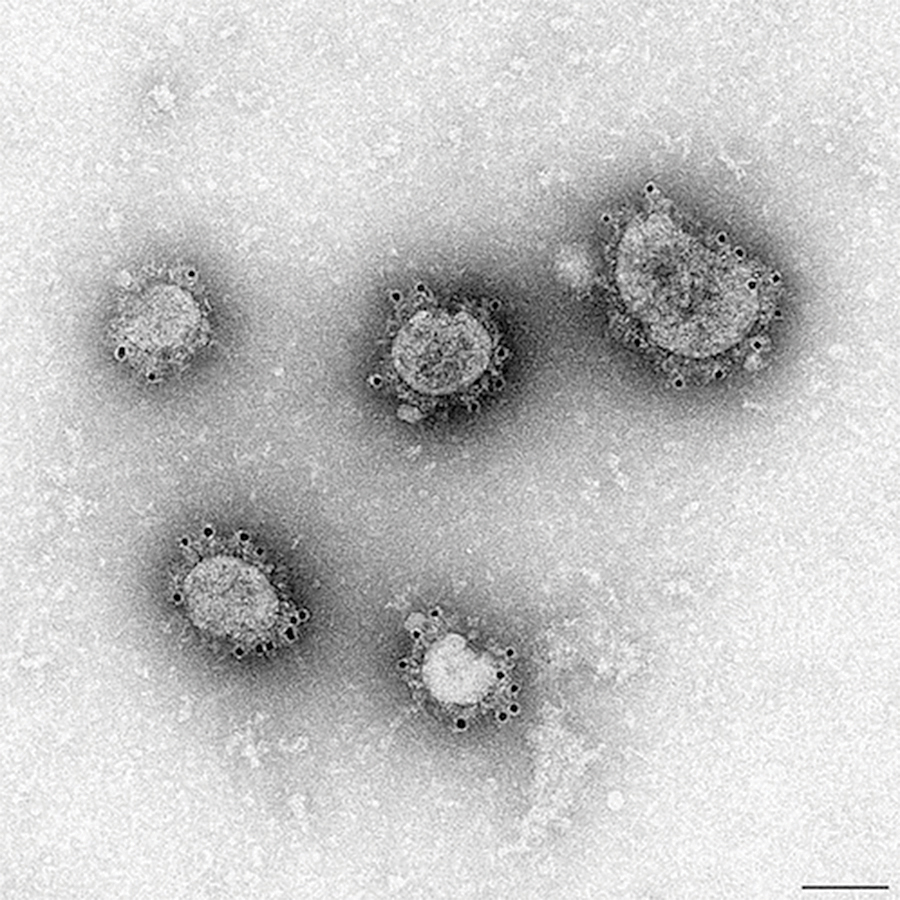
Immunogold labeling of Middle East Respiratory Syndrome-coronavirus
(MERS-CoV). MERS virus is grown in VERO cells via a grid cell culture technique. Primary antibody is a camel anti MERS serum which reacts with the glycoprotein spikes of the virus. Detection with Protein A 10nm. After incubation with the gold reagent specimens are fixed in glutaraldehyde and negative stained with Nano-W.
Courtesy of Sandra Crameri, Australian Animal Health laboratory CSIRO, Geelong, Australia
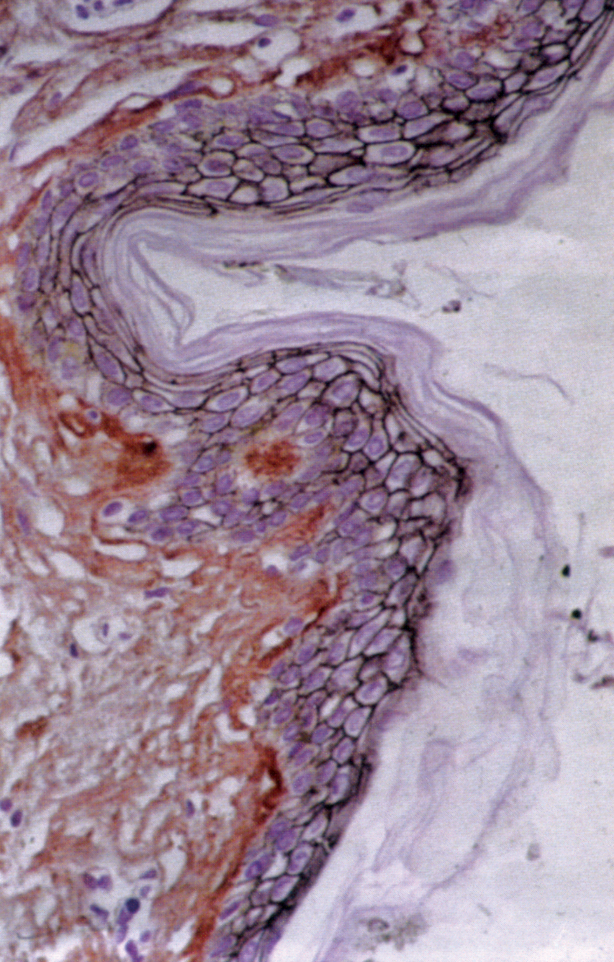
Immunogold Silver Staining of E-cadherin on a paraffin section of human skin. Courtesy of R. Moella, Dept. of Exp. Path., EUR, The Netherlands. • Mouse monoclonal anti E-cadherin • GAM lgG UltraSmall • Aurion R-Gent SE-LM
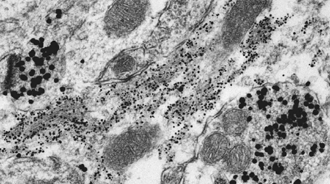
Pre-embedding double labeling of GFAP (small particles) and synaptophysin (large particles) in brain tissue using two ultrasmall gold conjugates and silver enhancement. H. Yi
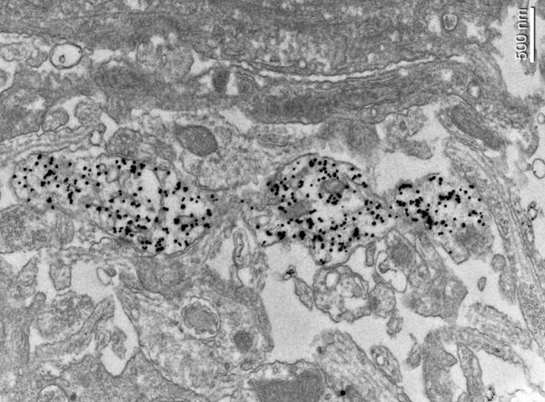
Pre-embedding labeling on 2%PFA/GA fixed and freeze thaw permeabilized organotypic cultured brain slices using rabbit anti GFP, AURION GAR 2nm and R-Gent SE-EM.
Courtesy of Y. Sun, D. Guerrero-Given and N. Kamasawa; EM Core Facility, Max Planck Florida, Institute for Neuroscience, Jupiter, FL USA
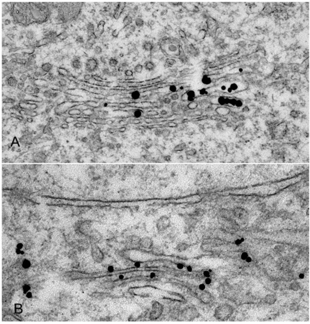
Pre-embedding labeling of MGP-160 (A) and HIPI protein (B) in brain tissue using ultrasmall gold conjugates and silver enhancement. Note the localization of the gold particles in the inner and outer surface of golgi membrane. H. Yi
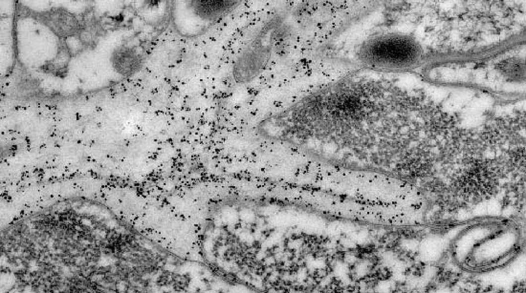
Post-embedding labeling of G12 in the symbiosome membrane using ultrasmall gold conjugate and silver enhancement.
T. Wakefield and H. Yi
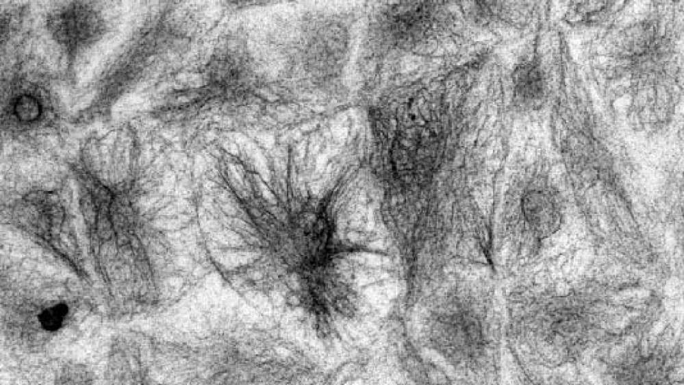
Pre-embedding labeling of vimentin in cultured microvascular endothelial cells using ultrasmall gold conjugate and silver enhancement. H. Yi